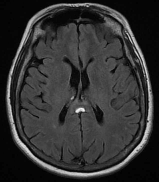
A middle aged woman was seen in the emergency department for confusion, and a non-enhanced head CT was obtained, shown below. Her exam was otherwise normal.
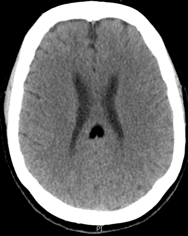
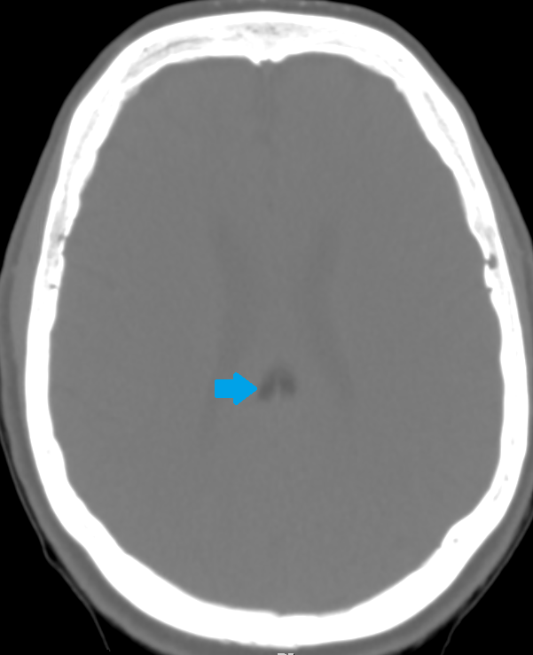
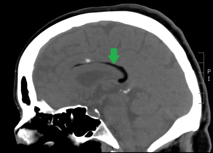
All three images above are non-enhanced head CT’s. First, locate the lesion – it is highlighted by a green arrow in the saggital, and by a blue arrow with bone windowing in the axial imaging. Where is it? What is the density compared to the surrounding brain tissue and CSF? Given the density, what should be in your differential diagnosis?
The most likely structure identified by the green and blue arrows here is:
- A) Free air
- B) Midline astrocytoma
- C) Lipoma
- D) Low grade brain abscess
Scroll down to see the answer.
The answer is B) this is a lipoma. Only fat and air have a density below that of CSF on head CT. Although both lipomas and free air can look similar on normal brain windowing, by adjusting the image settings to bone windowing (middle image with blue arrow above), you will see that the lipoma is no longer totally dark.
Lipomas are relatively common, benign fatty tumors. They tend to be in the midline, just like this one is. They are usually developmental and rarely cause problems later in life – most of the time they are incidental findings like in this case. If large, they can disrupt the development of midline structures like the corpus callosum.