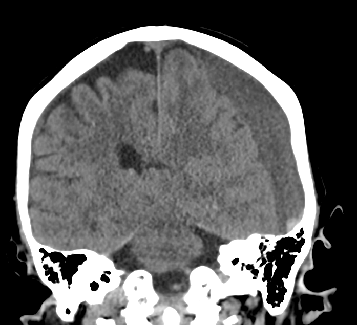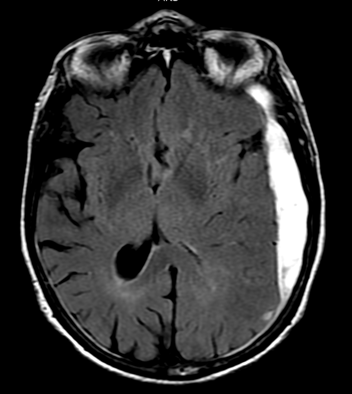
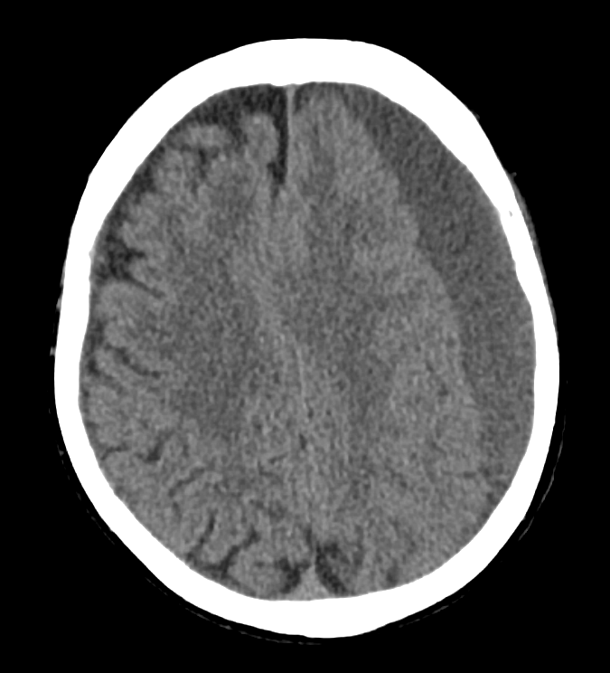
Subdural hematomas are collections of blood between the brain’s tough dural covering and thin arachnoid covering. The most common cause is tearing of venous structures, and it can be caused by minor trauma. Patient’s on anticoagulants, thrombocytopenia or significant cerebral atrophy are at higher risk.
Clues to the presence of subdural hemmorhages include:
- No extension into the sulci (as would be seen in subarachnoid bleeding)
- Concave shape, like a crescent
- Not typically associated with skull fractures, unlike epidural hematomas
- Typically slowly expanding