Interpreting and localizing visual field defects.
An elderly man presented to his ophthalmologist with complaints of difficulty seeing. Confrontational visual field testing revealed a defect, and formal visual field testing was done and is shown below.
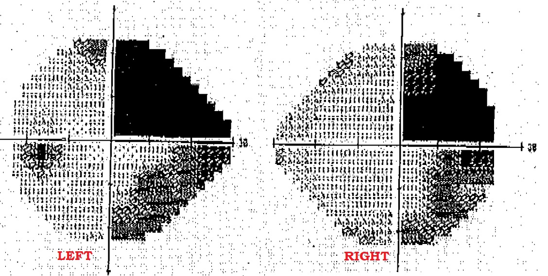
Let’s break down the visual field results above. First, it’s important to know that the typical bedside confrontational test (ie counting fingers), is a fairly crude test for vision loss. Formal tests include the Humphrey and Goldmann visual field testing, and require special equipment. They also require good patient cooperation.
The left eye is presented on the left side of the image, and the right eye is on the right side. Darker shading indicates vision loss. What type of vision loss does this patient have? A right homonymous superior quadrantopia. Also – do you see the physiologic blind spot in the visual fields above? (Hint: best seen in the left eye results).
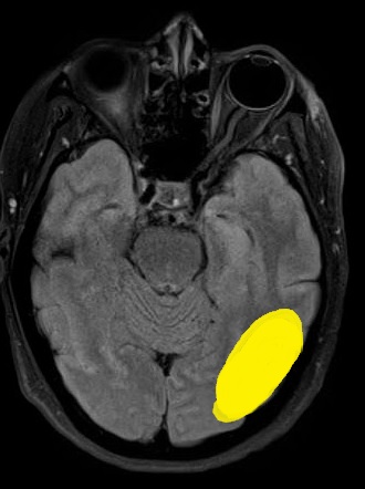
This superior wedge shaped area of vision loss is called a “pie in the sky.”
How do you localize a pie in the sky visual field defect? Well, the visual field defect affects both eyes, which tells you that it is behind the optic chiasm. Remember also that visual input is processed in the contralateral hemisphere, so a right sided visual defect localizes to the left hemisphere. Lastly, visual processing is inverted on the vertical (superior-inferior) axis.
A right homonymous superior quadrantopia localizes to a left temporal or occipital lobe lesion. Recall that the optic radiations pass through both the posterior temporal and parietal lobes.