
A few pointers from a single illustrative head CT.
Lets take a look at the head CT from a single patient, admitted to the neuro ICU three days ago. Can you guess from the image what the admitting diagnosis is?
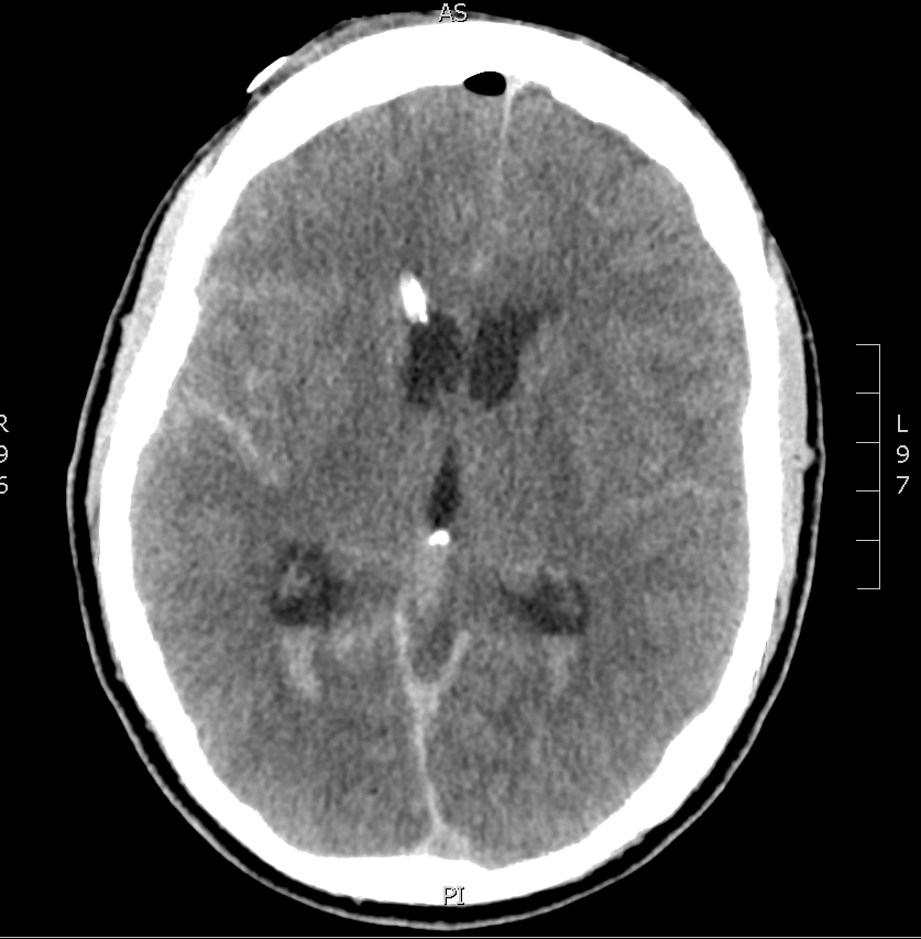
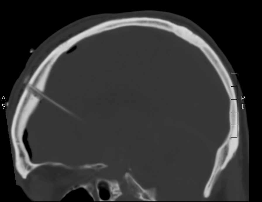
There is a lot going on in this CT – but you may have realized that there is diffuse subarachnoid hemorrhage. There are a number of other findings that we’ll walk through. First, take a look at using our coronal and sagittal views – fun fact, these are simply computer reformats of the axial above.

Coronal CT 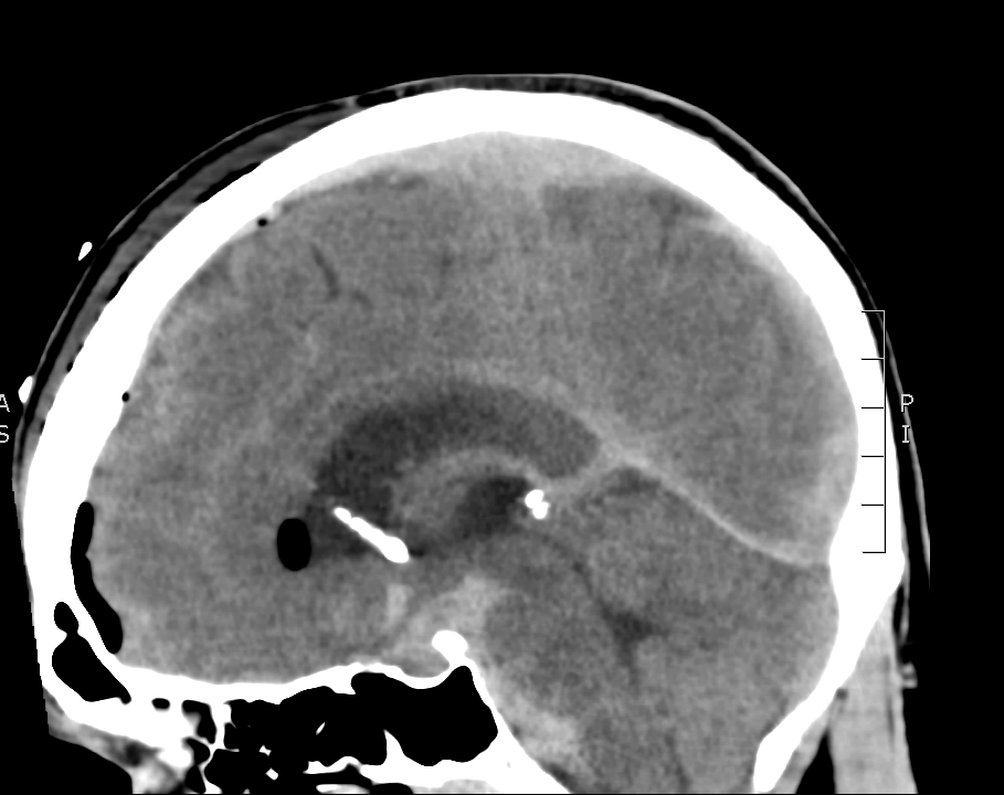
Sagittal CT
Now, let’s get into the specifics of the axial image. Scroll down for the key.
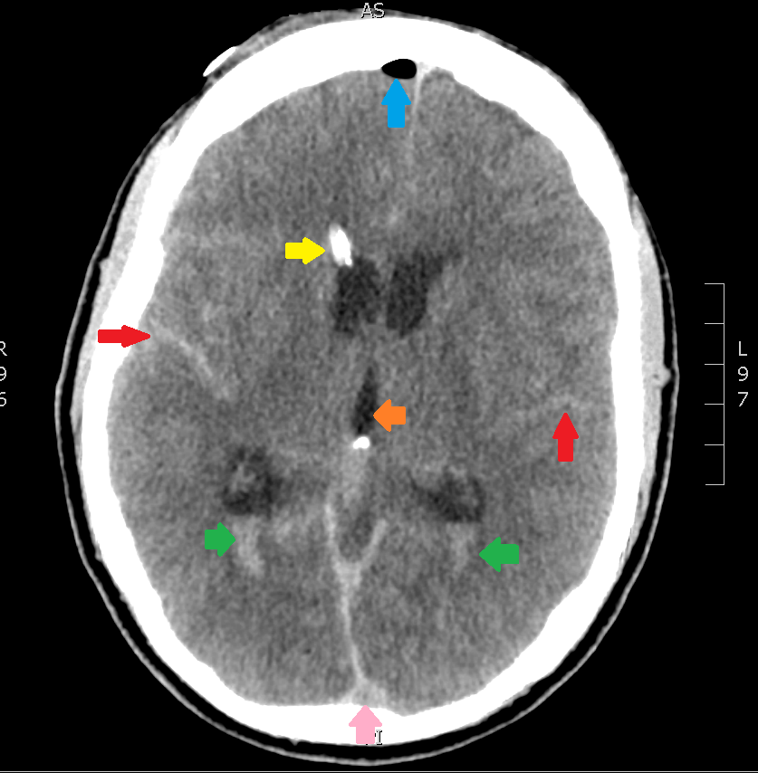
The highlighted structures are:
- Red Arrow – subarachnoid blood; you know it’s blood because it’s hyperdense, and it’s subarachnoid because it’s settled inside the cerebral gyri.
- Green Arrows – this is intraventricular blood, layering out in the bottoms of the lateral ventricles due to gravity (in this case, the source is the subarachnoid blood).
- Yellow Arrow – a tricky one, this is a ventricular drain, or EVD – commonly used to prevent the development of hydrocephalus after subarachnoid hemorrhage. Look for it again in the coronal and sagittal images above.
- Blue Arrow – Free air, given how hypodense it is, and it has found its way to the top of the cranium, as you would expect an air bubble to do. The source if the EVD.
- Orange Arrow – The third ventricle
- Pink Arrow – Dural venous sinus
If you go back to the coronal and sagittal images you can try to find the same structures. You’ll also see that there is a fair amount of subarachnoid hemorrhage anterior to the brain stem.
There can be a lot of information in a single head CT. For more details in interpreting neuro-imaging download the PDF of the free Neuroimaging e-book.

1 Comment