
How to identify pneumocephalus on a head CT.
Air occasionally enters the cranium and fills the ventricles – usually after a neurosurgical procedure, severe head trauma, or ventriculostomy placement. Air inside the skull is called pneumocephalus.
Although high flow oxygen can be used to help speed up the absorption of free air, but post-surgical pneumocephalus tends to be asymptomatic and resolves spontaneously. Take a look at the axial head CT below – do you see the free air? Scroll to the bottom for an annotated image.
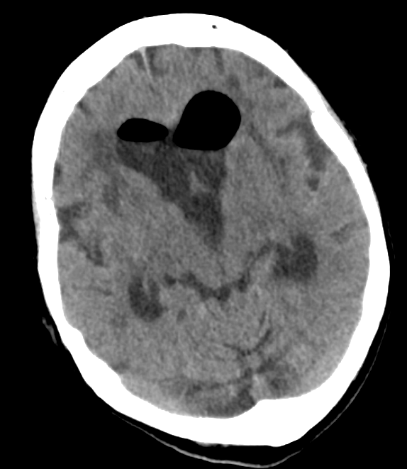
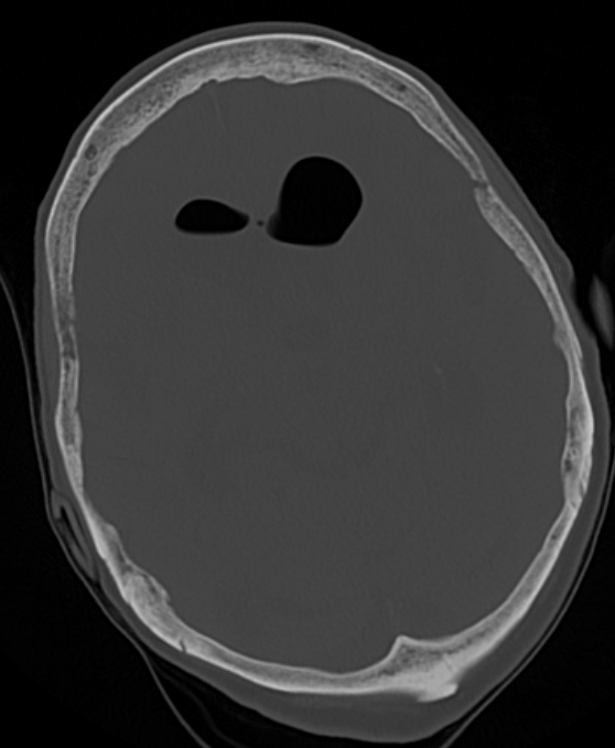
Air has an extremely low density on head CT, and should appear black, no matter how you window the head CT. One of the easiest ways to verify that something dark is air, rather than another hypodense material such as fat, is to look at the CT using the bone window, a setting which shows detail in highly dense structures. Air should remain dark even when viewing the head CT in this way – see the example below.
Did you see the free air? Look at the annotated head CT below to be sure.

Rarely, arterial air emboli can cause strokes.
For more on head CT’s, see the blog post on CT basics. Alternatively, check out the free neuro-imaging e-book.
