
How to identify Wallerian degeneration on imaging
Wallerian degeneration is the process of retrograde degeneration of nerve axons following an injury that separates them from the cell body of the nerve. This happens because the axon no longer receives proteins and nutrients from the cell body. It occurs in both the peripheral and central nervous system.
Scroll through the T2 FLAIR images in the slideshow below to follow a left basal ganglia injury with Wallerian degeneration of the descending motor axons in the brain stem.

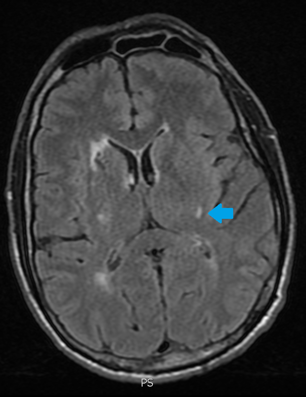


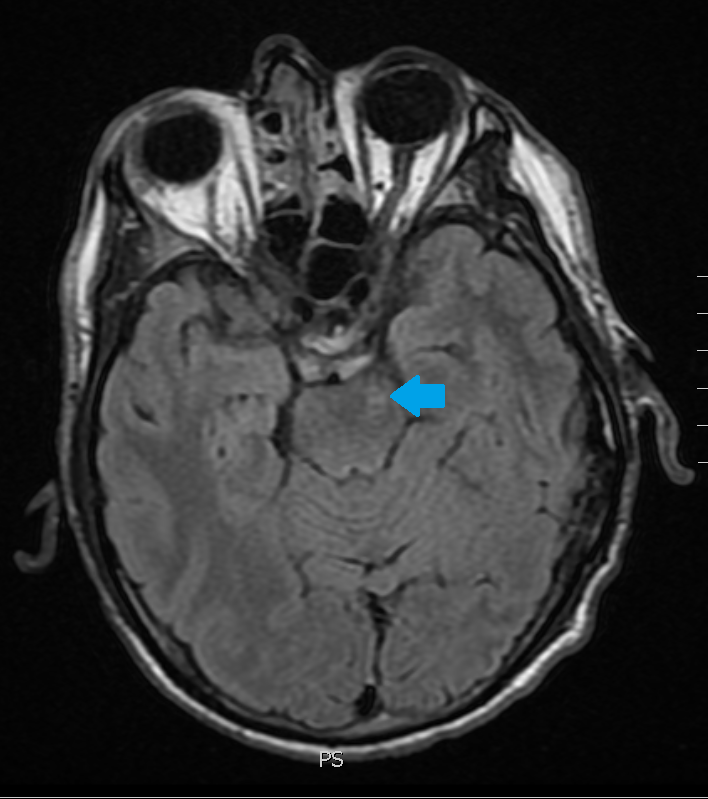
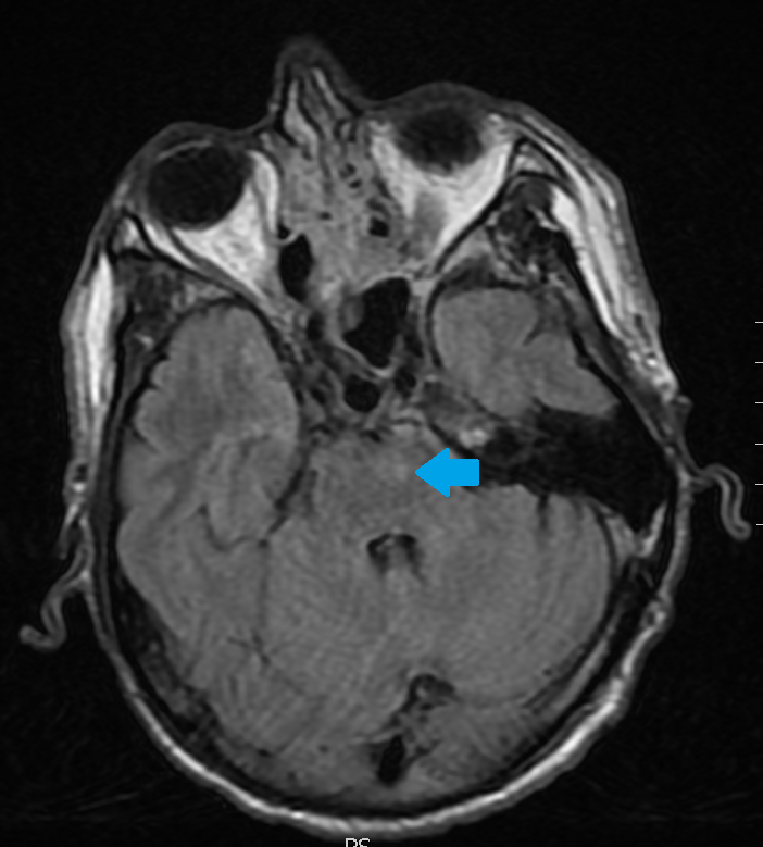
The original injury was the small lesion on the first slide. The white matter abnormality pointed out on the subsequent slides is from Wallerian axonal degeneration.
How quickly do the axons of injured nerves regenerate? (scroll down for the answer)
- A) 1 cm/day
- B) 1 cm/week
- C) 1 mm/day
- D) 1 mm/week
- E) 1 um/day
Answer: Nerve axons regenerate at a rate of 1 mm/day.