
A US major general and personal physician to Presidents Cleveland and McKinley is found to have a brain mass.
Major General Leonard Wood (1860 – 1927) was a physician, US Army Chief of Staff and Military Governor of Cuba. He was also one of the first individuals to have a brain tumor surgically removed; a procedure performed by Harvey Cushing, the father of neurosurgery, in 1910.
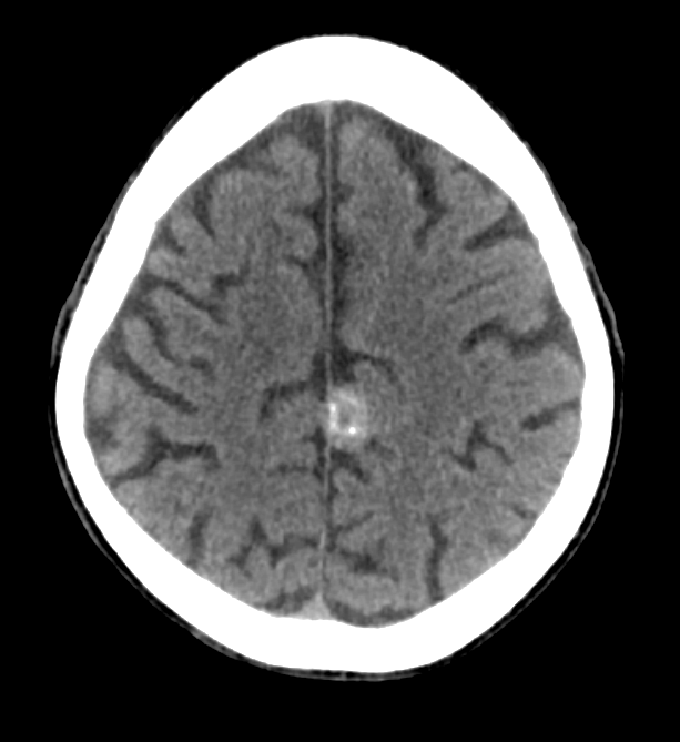
See below for a representative head CT.
Wood’s symptoms had included progressive headaches, weight gain, and personality changes.
Based on the CT scan shown above, which of the following are true? (scroll down for the answer)
- The lesion is EXTRA-AXIAL
- The lesion is INTRA-AXIAL
- The lesion is MALIGNANT
- The lesion is HEMORRHAGIC
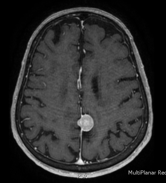
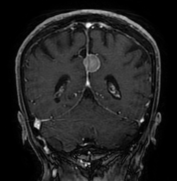
The answer is 2) the lesion is extra-axial. Take a look at the brain MRI’s below to prove it to yourself.
Now, which of the following is true? (scroll down for the answer)
- The lesion contains psammoma bodies
- The lesion contains rossettes
- The lesion contains Tau
- The lesion contains rosenthal fibers
The answer is 1) these lesions often contain small bits of calcium, called psammoma bodies. You have probably guessed by now that General Wood had a meningioma – a brain tumor that is usually benign, although they can cause significant symptoms depending on their location and degree of mass effect. Meningioma’s rarely recur after surgical resection, although unfortunately for General Wood, his did.
Meningiomas should be broad based to the dura, as is the example shown here, which grows out of the falx cerebri. Most are sporadic, but exposure to ionizing radiation is a risk factor for late life meningiomas.

