
Learn how to quickly identify acute stroke on MRI.
If you haven’t already read the post on T1 and T2 weighted sequences on the brain MRI, check it out here.
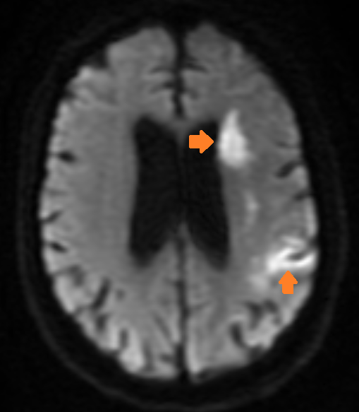
Brain MRI is very sensitive for acute stroke, making it an extremely useful tool for the neurologist. The most accurate MRI sequence for identifying acute stroke is called Diffusion Weighted Imaging (DWI).
On a DWI sequence, acute stroke will appear bright, making it easy to quickly identify.
The DWI signal, called diffusion restriction, is usually apparent within the first hour of stroke symptom onset, although very early MRI may miss small brain stem strokes. DWI stays bright for about two weeks, before normalizing. Occasionally, the DWI signal will persist long after the acute stroke – a phenomenon called “shine through.”
To avoid mistaking DWI shine through for acute stroke, neurologists and radiologists refer to a second MRI sequence, called the ADC map. This ‘confirms’ acute stroke by appearing dark in the corresponding area of diffusion restriction. The ADC map is dark for about two weeks. If the diffusion is bright, and ADC is not dark, then you’re probably looking at shine through, e.g. a stroke older than 2 weeks – possibly much older.

Not everything that shines brightly on diffusion is a stroke – there are a few mimics. Some hyperdense tumors, brain abscesses, and in rare cases, the edges of demyelinating lesions, can have diffusion restriction.
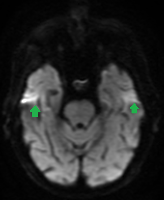
In addition, there are common artifacts on DWI, especially at the temporal poles, were aberrant signal is often present – see below for an example.

1 Comment