
An introduction to T1 and T2 Weighted imaging in MRI.
Understanding the difference between T1 weighted and T2 weighted sequences on a brain MRI is a very useful skill, as these are two of the most common sequences. Each sequence in an MRI is obtained independently. T1 vs T2 refers to the timing and physics of how the sequence is obtained.
T1 Weighted Sequences
T1W sequences are the best for showing brain anatomy.
- White matter looks white
- Grey matter looks grey
- CSF is dark
- Fat is bright
- A few other items are ‘bright’, such as gadolineum contrast and some mineralization

T2 Weighted Sequences
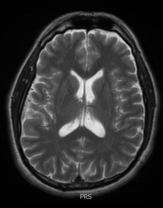
T2W sequences show water, and therefore inflammation and pathology, – T2 shows bright H2O
- White matter looks grey
- Grey matter looks white
- CSF is BRIGHT
- Fat is bright (just like in T1)
- Because CSF is bright, it can be hard to see pathological edema
T2 FLAIR Sequences
T2 FLAIR shows the pathological edema best highlighted in T2 sequences, but removes the bright CSF signal, making pathology ‘pop out’. This is a go-to, work horse sequence on MRI. Be aware that hyperacute strokes may no cause a FLAIR change.
- White matter looks grey
- Grey matter looks white
- CSF is DARK
- Fat is bright (just like in T1)
- Highlights pathology

OK – time to review, can you tell which sequence is T1W and which is T2W? Look at the two saggital MRI’s below and decide which is which, then scroll down for the answer.
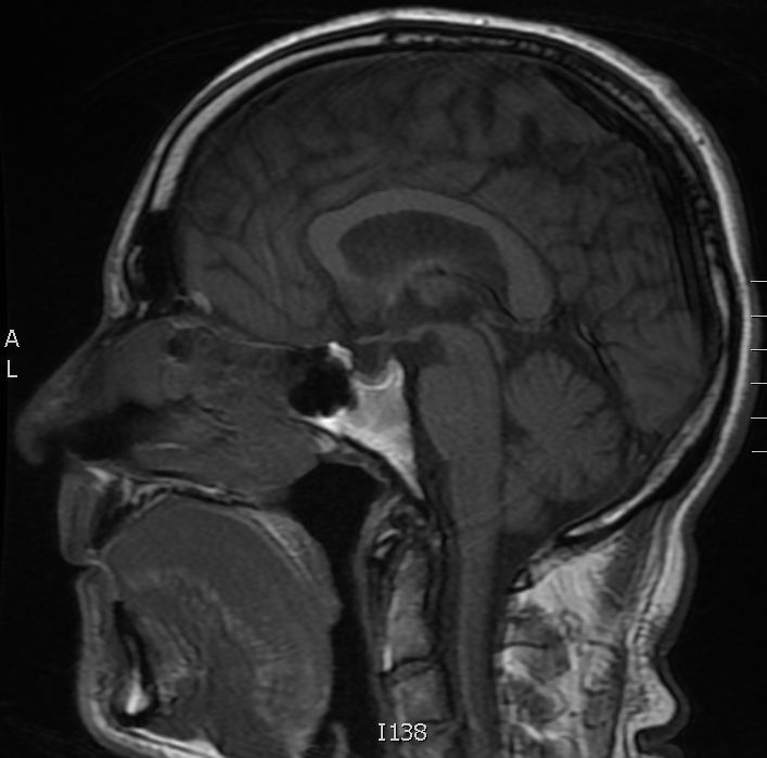
Image A 
Image B
Image A is a T1W study – the corpus callosum, a large white matter tract, is bright. Image B is a T2W FLAIR study, the corpus callosum is grey. Notice that the fat is bright on both.
For more information on MRI sequences, download our free neuroradiology guide! To learn about diffusion restriction and acute strokes, read this post.

5 Comments