What’s on the differential diagnosis for a ring enhancing lesion?
The term ‘ring enhancing’ refers to lesions which develop a circular, doughnut-like rim of enhancement after contrast administration. Hint: only post-contrast studies can show ring enhancing lesions.
There is a useful mnemonic for differential diagnosis of ring enhancing lesions- “Magical Doctor”:
M A G I C L DR

Pre-contrast T1W MRI 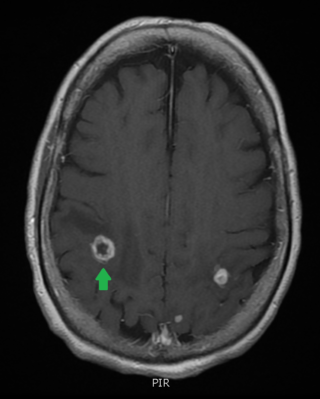
Post-contrast T1W MRI
First, lets take a look at the importance of using contrast in detecting lesions – the MRI on the left is pre-contrast. You can see a circular lesion (maroon arrow), but with contrast administration (image on the right) you now see a true “ring enhanced lesion” that is obvious and well defined, but you also notice two additional lesions on the left hemisphere (not marked).
So what might cause these ring enhancing lesions? M A G I C L DR will help us remember the differential diagnosis.
Malignancy – most often metastatic disease
Abscess – usually with surrounding edema
Glioblastoma – the classic form can cross the corpus callosum and have a “butterfly” appearance
Infarct – subacute strokes can show peripheral enhancement; not thought of as frequently because stroke patients rarely receive contrast enhanced studies in the subacute stroke phase.
Contusion – a resolving parenchymal hematoma may show contrast enhancement
Lymphoma
Demyelination – occasionally you will see a rim of enhancement in actively demylinating lesions; typically an C – shaped, open rim of enhancement
Radiation – less common nowadays, but radiation injuries could do this


Above: More examples of ring enhancing lesions (blue arrows). Note multiple other enhancing lesions scattered throughout.
In this case, the patient was diagnosed with CNS lymphoma – he was chronically immunocompromised due to a history of solid organ transplant.

From now on, every time you eat a doughnut, take the opportunity to review MAGICL DR 😛

1 Comment