
The butterfly lesion – learn how to recognize a glioblastoma multiforme (GBM) on MRI.
We recently published a post about ring-enhancing lesions, using the mnemonic MAGICL DR to remember the differential diagnosis.
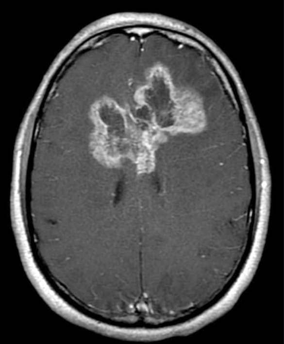
One important ring enhancing lesion on brain MRI is a glioblastoma multiforme, or GBM – an aggressive type of brain tumor. A GBM is a high grade primary brain tumor. It develops from atrocytes, and is therefore called a stage IV astrocytoma.
Glioblastomas have a tendency to spread across the corpus callosum, creating a bihemispheric lesion, sometimes described as resembling a butterfly.

GBM’s represent about 15% of all intracranial tumors, and are the most common adult intra-axial tumor, which refers to tumors arising from within the brain. (Reminder, the most common extra-axial tumor is a meningioma).
The inner core of a GBM represents necrotic tissue. The outer, enhancing ring is made of hyper-vascularized tissue. GBM’s spread quickly, and are aggressive brain tumors with poor long term prognosis.


GBM’s often have a shaggy, contrast enhancing ring surrounding an area of central necrosis. There may be edema around the lesion. They may cross the corpus callosum, or extend into the brainstem.
Consider reviewing our post about MRI basics as well. Neuro-oncology is covered at length in the review books.
